Nociception
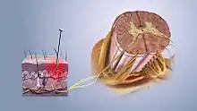
La nociception est une fonction défensive présente chez les vertébrés ainsi que chez les invertébrés[1]. C'est une fonction permettant l'intégration, au niveau du système nerveux central, d'un stimulus potentiellement nocif via l'activation des nocicepteurs cutanés, viscéraux, musculaires et articulaires[1]. Le transport de l'information sensorielle par les nerfs se fait de la périphérie (lieu de transduction d'un message potentiellement nocif en nociception) jusqu'au thalamus. La nociception est souvent décrite comme transmettant les messages "douloureux", cependant cette conception est erronée[1] - [2] - [3]. En effet la nociception et la douleur sont deux choses différentes [1] - [4]. Il existe un grand corpus de preuve dissociant la nociception de la douleur, montrant qu'il n'existe pas de lien de causalité entre le premier et le second [5] - [6] - [7] - [8].

Voir aussi
Articles connexes
Notes et références
- (en) W. Daniel Tracey, « Nociception », Current Biology, vol. 27, no 4, , R129–R133 (DOI 10.1016/j.cub.2017.01.037, lire en ligne, consulté le )
- G. Lorimer Moseley, Explain Pain, (ISBN 978-0-9873426-6-9 et 0-9873426-6-5, OCLC 861699508, lire en ligne)
- David S. Butler, Explain pain supercharged : The clinician's manual, (ISBN 978-0-6480227-0-1 et 0-6480227-0-6, OCLC 982125476, lire en ligne)
- (en-US) Srinivasa N. Raja, Daniel B. Carr, Milton Cohen et Nanna B. Finnerup, « The revised International Association for the Study of Pain definition of pain: concepts, challenges, and compromises », PAIN, vol. 161, no 9, , p. 1976–1982 (ISSN 0304-3959, PMID 32694387, PMCID PMC7680716, DOI 10.1097/j.pain.0000000000001939, lire en ligne, consulté le )
- (en) Teun Teunis, Bart Lubberts, Brian T. Reilly et David Ring, « A systematic review and pooled analysis of the prevalence of rotator cuff disease with increasing age », Journal of Shoulder and Elbow Surgery, vol. 23, no 12, , p. 1913–1921 (ISSN 1058-2746 et 1532-6500, PMID 25441568, DOI 10.1016/j.jse.2014.08.001, lire en ligne, consulté le )
- (en) Jonathan M. Frank, Joshua D. Harris, Brandon J. Erickson et William Slikker, « Prevalence of Femoroacetabular Impingement Imaging Findings in Asymptomatic Volunteers: A Systematic Review », Arthroscopy, vol. 31, no 6, , p. 1199–1204 (ISSN 0749-8063 et 1526-3231, PMID 25636988, DOI 10.1016/j.arthro.2014.11.042, lire en ligne, consulté le )
- (en) Adam G. Culvenor, Britt Elin Øiestad, Harvi F. Hart et Joshua J. Stefanik, « Prevalence of knee osteoarthritis features on magnetic resonance imaging in asymptomatic uninjured adults: a systematic review and meta-analysis », British Journal of Sports Medicine, vol. 53, no 20, , p. 1268–1278 (ISSN 0306-3674 et 1473-0480, PMID 29886437, PMCID PMC6837253, DOI 10.1136/bjsports-2018-099257, lire en ligne, consulté le )
- (en) W. Brinjikji, P. H. Luetmer, B. Comstock et B. W. Bresnahan, « Systematic Literature Review of Imaging Features of Spinal Degeneration in Asymptomatic Populations », American Journal of Neuroradiology, vol. 36, no 4, , p. 811–816 (ISSN 0195-6108 et 1936-959X, PMID 25430861, PMCID PMC4464797, DOI 10.3174/ajnr.A4173, lire en ligne, consulté le )